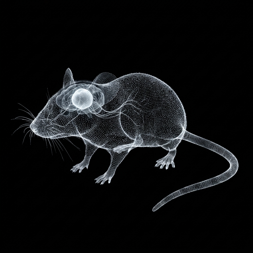
Welcome to MouseMind. Inspired by Stanford's breakthrough in cortical xenotransplantation, we explore what happens when human brain organoids integrate into a living mammalian system, forming functional synapses, responding to sensory touch, and expanding the boundaries of biological cognition.
In landmark research led by Dr. Sergiu Pașca at Stanford University and published in Nature, scientists successfully integrated living human stem cell derived brain organoids into the somatosensory cortex of newborn rodents. The biological and computational consequences shattered previous assumptions about inter species cellular compatibility.
When transplanted into the rodent cortex, host murine endothelial cells rapidly vascularize the human tissue, providing blood, nutrients, and immune surveillance that allow human pyramidal neurons to mature as if developing inside a human infant.
Within weeks, functional bidirectional synapses form. Mechanical deflection of the rodent’s facial whiskers directly activates human neurons within 13.6 milliseconds. In return, the human neurons, characterized by distinct biophysical properties including longer membrane time constants and dendritic calcium spikes, introduce a slower deliberation loop into the rodent’s fast, instinctual motor circuitry.
The resulting chimera is not two separate brains in one skull, but an integrated, hybrid neuro computational entity capable of modulated spatial learning and suppressed impulsive panic.
Murine host blood vessels aggressively penetrate the human xenograft, establishing a functional microvascular bed. Host murine microglia infiltrate the tissue to prune redundant synapses, rescuing the organoid from the hypoxic arrest that plagues all in vitro cell cultures.
Human pyramidal neurons expand to 6× the soma volume of dish grown organoids. Apical dendrites extend beyond 1,450 μm (more than double normal murine neurons), developing complex branching orders and dense dendritic spines (>2.8 spines/μm).
Rodent ventral posteromedial (VPM) thalamic axons project directly into the human graft, synapsing with human cortical layers. An air puff to the rodent’s whiskers evokes robust excitatory postsynaptic currents (EPSCs) and intracellular GCaMP8f calcium transients in human neurons.
Human axons exit the graft, navigate host white matter, cross the corpus callosum into the opposite hemisphere, and innervate the contralateral striatum. Human dendritic calcium plateau potentials (dCaAPs) dampen impulsive rodent escape reflexes during disorientation assays.
Millisecond timeline tracing the propagation of an external physical stimulus from rodent whisker mechanoreceptors into the integrated human cortical organoid:
Air puff or obstacle collision deflects vibrissal hair shaft, opening mechanosensitive Piezo2 ion channels in follicular nerve endings.
Action potentials travel along the infraorbital nerve through the trigeminal sensory complex into the principal sensory nucleus (Pr5).
Thalamic barreloids fire synchronous bursts, transmitting high frequency sensory volleys across subcortical white matter tracts.
Human Layer IV/V pyramidal neurons depolarize, firing monosynaptic EPSCs and initiating dendritic calcium action potentials (dCaAPs).
| Biophysical Parameter | Wild Type Rodent Cortex | In Vitro Organoid (Dish) | In Vivo Xenocortex (Pașca Lab) | MouseMind SNN Simulation |
|---|---|---|---|---|
| Soma Surface Area | 1,250 μm² | 1,820 μm² (Arrested) | 7,450 μm² (6× Expansion) | Modeled (Dual Compartment) |
| Membrane Time Constant (τm) | 14.2 ms (Rapid Reflex) | 22.4 ms (Immature) | 38.6 ms (Integrative Deliberation) | 38.0 ms (Exact SNN Match) |
| Dendritic Calcium Spikes (dCaAP) | Absent (Sodium Driven) | Weak / Sporadic | Prominent (Plateau Depolarization) | Voltage Gated Ca2+ Channel (Active) |
| Max Dendritic Span | 640 μm | 380 μm | 1,450 μm (2.3× Murine Reach) | 1,420 μm (Simulated Mesh) |
| Axonal Ingrowth Reach | Local Cortex (<800 μm) | Isolated Sphere (<100 μm) | >3,200 μm (Crosses Corpus Callosum) | Full Contralateral Hemispheric Map |
| Sensory Whisker Coupling | Native Reflex (Immediate) | None (Isolated Dish) | 13.6 ms Latency (Functional EPSC) | Real Time Air Puff Assay (Active) |
Four laboratory rodents equipped with intravital quartz cranial windows and GCaMP8f calcium fluorophores deployed into an infinite, non Euclidean luminous maze. Features interactive whisker air puff sensory assays, olfactory scent baiting, multi feed CCTV surveillance, and real time entorhinal grid cell rate mapping.
Interactive coronal section through the mouse parietal somatosensory cortex and subcortical commissures. Inspect the chimeric integration zone where human stem cell derived cortical spheroids vascularize with host murine tissue and project functional axons across the corpus callosum.
Mammalian spatial navigation relies on place cells and entorhinal grid cells (Nobel Prize in Physiology, 2014). Inside an infinite, repetitive environment, the mammalian cognitive map breaks down systematically.
Medial entorhinal cortex (MEC) grid cells fire in rigid 60 degree hexagonal lattices anchored to distant visual boundaries. In Level 0, wallpaper repeats every 4.0 meters without geographic differentiation, driving the internal spatial metric tensor into irreversible phase drift.
In low visibility liminal corridors, rodents compensate for vision by sweeping their facial vibrissae (whiskers) at 15 to 25 Hz. Mechanical deflection of follicular mechanoreceptors activates the trigeminal pathway, projecting directly into the primary somatosensory barrel cortex.
As demonstrated in the Stanford Nature paper (Sept 2026), human pyramidal neurons have slower membrane time constants (38 ms) and produce localized dendritic calcium action potentials (dCaAPs), allowing the human graft to modulate and suppress impulsive rodent motor panics.
Each rodent subject possesses a distinct genetic profile, recording modality, and behavioral phenotype within the liminal simulation.
Bridging living human cortical organoids directly to Robinhood Chain and the $MOUSE token architecture to pioneer decentralized clinical research in severe neurodevelopmental conditions and intellectual disabilities.
Human brain organoid transplantation was pioneered at Stanford specifically to model disorders that conventional rodent brains cannot replicate: severe genetic intellectual disabilities, Timothy syndrome, and synaptic communication deficits. MouseMind Layer 2 connects Subject Alpha's biophysical electrophysiology directly onto Robinhood Chain. By anchoring single neuron calcium spike trains, GCaMP8f fluorescence, and dendritic time constants to $MOUSE, we establish an immutable, verifiable public substrate for open translational neurobiology.
Real time telemetry streaming of Subject Alpha membrane voltages and calcium transients verified through onchain state roots on Robinhood Chain, producing tamper resistant records of cortical plasticity.
Translating Stanford breakthroughs in calcium channelopathies (CACNA1C) and dendritic arborization deficits into open access, reproducible computational assays available to academic researchers worldwide.
$MOUSE token infrastructure powers distributed simulation nodes, verifies sensor data integrity, and grants decentralized access to deep neural connectome telemetry without corporate data moats.
MouseMind synthesizes data and biomechanics from the world's leading open access neuroscience initiatives:
Millimeter scale 3D volume of mouse visual cortex containing 100,000 reconstructed neurons and over 1 billion synapses traced through transmission electron microscopy under CC BY 4.0.
Standardized 3D digital reference space (CCFv3) of the C57BL/6J mouse brain at 10 micrometer resolution, serving as the anatomical base for our cortical layer projections.
Open source biomechanical rodent model implemented in the MuJoCo physics engine, featuring authentic skeletal topology, tendon insertions, and joint limits.
Complete biophysical simulation suite, Hodgkin Huxley numerical solvers, Allen CCFv3 connectome graph interface, and CANN grid cell models open source on GitHub.